
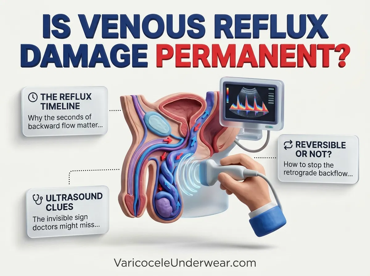
Looking at a scrotal ultrasound printout can be confusing, especially when it contains terms such as retrograde flow, venous diameter, and Valsalva maneuver. For men diagnosed with a varicocele, understanding varicocele venous reflux duration is particularly important. This measurement describes how long blood flows backward through the enlarged testicular veins during testing. Current European urological guidance uses a reflux duration greater than two seconds, measured while standing during a Valsalva maneuver, as an important indicator of a clinically significant varicocele. However, this number must be interpreted alongside vein diameter, testicular volume, physical examination findings, and symptoms.
Decoding Your Scrotal Ultrasound Report
A scrotal Doppler ultrasound combines standard imaging with Doppler technology, which measures the direction and characteristics of blood flow. The examination focuses on the pampiniform plexus, a network of veins that carries blood away from the testicles. When the valves inside these veins do not work correctly, blood can move backward and collect in the scrotal veins. This condition is called a varicocele. When reading a scrotal Doppler report, look for several specific findings: the largest measured vein diameter, the presence and duration of retrograde flow, the patient’s examination position, and the testicular volume on each side. You can also review our guide to interpreting Doppler ultrasound varicocele imaging results for a closer explanation of common report terminology.
The examination technique matters because venous pressure changes with body position and abdominal pressure. A standardized assessment generally includes imaging both testicles while the patient is lying down and standing. The sonographer evaluates blood flow during normal breathing and during the Valsalva maneuver, which involves briefly bearing down as though trying to have a bowel movement. This action increases abdominal pressure and may reveal backward blood flow that is less apparent at rest. Color Doppler displays the direction of blood movement, while spectral Doppler provides a waveform that helps measure how long reversal lasts. Red and blue do not automatically mean arteries and veins; the colors represent flow direction relative to the ultrasound probe and the machine’s settings. A complete report should explain how and where measurements were taken rather than presenting numbers without context.
Understanding Retrograde Flow Duration
Varicocele Venous Reflux Duration: Understanding the Two-Second Threshold
Retrograde flow means blood moves in the opposite direction from its normal venous return toward the heart. During a Doppler examination, the radiologist measures how long this backward movement continues, particularly when the patient stands and performs the Valsalva maneuver. The European Association of Urology (EAU) identifies venous reflux lasting more than two seconds under these conditions as a finding associated with a clinically significant varicocele. The European Society of Urogenital Radiology Scrotal and Penile Imaging Working Group (ESUR-SPIWG) uses the same general threshold in its ultrasound recommendations. This measurement is important because prolonged reversal suggests abnormal venous valve function. It is not, however, a stopwatch measuring how long every drop of blood remains trapped in the scrotum. It describes the duration of the observed backward-flow episode during the examination.

Discover specialized varicocele underwear designed for daily comfort, support, and recovery — a more targeted option for men who want support without compromise.
Discover the Recommended OptionConsider two hypothetical Doppler results. One patient has reflux lasting 0.8 seconds during the standing Valsalva test, while another has continuous backward flow lasting 3.2 seconds. The second finding exceeds the commonly used diagnostic threshold and deserves clinical attention in the appropriate setting. A result of 1.5 seconds does not meet the greater-than-two-second criterion, but it cannot independently rule out a varicocele. Reflux may vary with positioning, the strength of the Valsalva maneuver, the measurement location, and ultrasound equipment settings. The radiologist should use spectral Doppler to confirm the flow pattern rather than relying on color images alone. Peak reflux velocity may also appear in a report, but ESUR-SPIWG identifies reflux duration as the essential parameter to measure. Velocity alone does not establish whether a patient needs treatment.
Another important distinction is the difference between an imaging finding and a treatment decision. A prolonged reflux measurement does not automatically mean that surgery is necessary, nor does a shorter measurement guarantee that fertility is unaffected. Some men have enlarged veins without pain or abnormal semen results. Others experience a dull ache, a dragging sensation, or discomfort after standing for several hours. The clinical examination remains central to diagnosis. Doctors also consider fertility goals, semen analysis, testicular size, and the severity of symptoms. Men concerned about persistent or recurrent reflux after a previous procedure should discuss whether repeat Doppler imaging is appropriate. Our guide to MRI versus ultrasound for varicocele recurrence explains why Doppler ultrasound is generally the first imaging choice when clinicians need to assess blood flow after treatment.
What Millimeter Vein Diameters Mean
Vein diameter is the second major measurement to examine when interpreting a varicocele ultrasound. The pampiniform plexus normally contains several small veins surrounding the spermatic cord. With a varicocele, these veins become enlarged and may appear as multiple winding, fluid-filled structures around the testicle. ESUR-SPIWG recommends measuring the largest vein while the patient is standing and performing the Valsalva maneuver. A maximum diameter of 3 millimeters or greater can be considered diagnostic under this standardized technique. The EAU describes a maximum diameter greater than 3 millimeters in the same position as a finding that correlates with a clinically significant varicocele. These small differences in wording reflect the use of specific guideline criteria, not a reason to interpret every measurement independently. A diameter measured while lying down or without Valsalva should not be compared directly with a measurement taken under the recommended conditions.
Ultrasound Diagnostic Parameters Reference
| Ultrasound parameter | Reference finding | Clinical meaning |
|---|---|---|
| Maximum vein diameter | 3 mm or greater under ESUR criteria | Supports a varicocele diagnosis when measured standing during Valsalva. |
| Venous reflux duration | Greater than 2 seconds | Abnormal prolonged backward flow under the standardized standing Valsalva test. |
| Examination position | Supine and standing | Helps reveal position-dependent venous enlargement and reflux. |
| Valsalva maneuver | Flow assessed during bearing down | Increases abdominal pressure to reveal abnormal venous reversal. |
| Spectral Doppler | Waveform and reflux duration recorded | Confirms flow direction and measures the duration of reversal. |
| Testicular volume | Both testicles measured | Helps identify size differences and possible testicular growth or tissue changes. |
Testicular volume deserves particular attention when fertility or testicular development is a concern. A smaller testicle may suggest reduced growth or atrophy, meaning a loss of testicular tissue volume. The EAU recommends calculating testicular volume using Lambert’s formula: length multiplied by width multiplied by height, then multiplied by 0.71. Comparing both sides gives the clinician useful information that vein diameter alone cannot provide. A man with a 3.5 mm vein and prolonged reflux, for example, may have a different clinical picture from someone with similar measurements but normal testicular volume, no discomfort, and normal semen parameters. Neither situation should be assessed using ultrasound measurements alone. For additional information about everyday support choices, see our guide to choosing supportive underwear for varicocele.
Understanding these measurements also helps translate an ultrasound report into practical daily management. Supportive underwear with gentle scrotal elevation may reduce movement, pulling, and the heavy sensation that some men experience during prolonged standing, walking, or exercise. VaricoceleUnderwear™ is designed to provide passive testicular support, which may improve physical comfort for some wearers. However, there is an important clinical distinction: mechanical elevation is not proven to shorten Doppler-measured retrograde flow duration, repair damaged venous valves, or eliminate a varicocele. The phrase ultrasound-tested varicocele uplift underwear should not be interpreted as evidence of a published clinical trial demonstrating shorter reflux duration. A supportive garment is a comfort measure, not a substitute for medical assessment or treatment. Its fit should feel supportive rather than tight or restrictive.
Finally, discuss your findings with a urologist, particularly if the report describes reflux lasting more than two seconds, veins measuring around or above 3 mm, reduced testicular volume, or persistent symptoms. Men experiencing fertility difficulties may also need semen analysis because ultrasound cannot determine sperm count, movement, or shape. The EAU recommends considering varicocele repair in appropriately selected infertile men who have a palpable clinical varicocele and abnormal semen parameters. Treatment decisions should account for the complete reproductive assessment rather than an isolated Doppler number. Sudden, severe testicular pain, rapid swelling, or a new firm lump requires prompt medical assessment because these symptoms may indicate conditions other than varicocele. For most men, a properly performed ultrasound combined with a clinical examination provides a useful starting point. Understanding the numbers can reduce unnecessary worry and help you ask better questions about monitoring, fertility, and treatment.
Clinical Evidence and Radiological Standards
The measurements discussed in this article follow published European urological and radiological recommendations. The ESUR-SPIWG systematic review by Bertolotto and colleagues, published in the Journal of Ultrasound in 2020, explains the standardized examination technique, the importance of spectral Doppler, and the two-second reflux threshold. Its recommendations are available in the European radiological standards for varicocele ultrasound assessment. These criteria help standardize imaging reports, but the final diagnosis and management plan must always account for the individual patient’s examination, symptoms, and reproductive health.
Related Articles



Choose Varicocele Support Designed for Cooling, Lift, and Daily Comfort
You’ve seen why regular underwear often falls short. Now view the purpose-built option made to reduce heaviness, improve support, and feel comfortable enough for everyday wear.