

Reading Your Scrotal Doppler Ultrasound Report
A scrotal duplex scan uses high-frequency sound waves combined with color Doppler technology to assess both anatomical structure and real-time blood movement. During the examination, an ultrasound probe evaluates the pampiniform plexus, a network of small veins responsible for draining deoxygenated blood away from the testicles. Established scrotal ultrasound reporting standards require the sonographer to record measurements in two distinct patient positions: lying flat on your back (supine) and standing upright. Gravity naturally pulls fluid downward, so standing measurements often reveal vascular enlargement that remains hidden while you are resting horizontally.The report usually documents both the left and right hemiscrotum separately. Left-sided varicoceles appear far more frequently because the left internal spermatic vein travels vertically into the left renal vein at a strict ninety-degree angle, generating higher hydrostatic pressure. The right spermatic vein drains at an angle directly into the high-volume inferior vena cava, encountering less natural resistance. If your diagnostic sheet describes isolated right-sided enlargement or sudden bilateral dilation, your doctor will check your abdominal anatomy to rule out secondary vascular compression.Millimeter Vein Diameters and Clinical Cutoffs
Vein diameter serves as the primary diagnostic yardstick on any scrotal imaging readout. In healthy men, pampiniform veins measure between 1.5 and 2.0 millimeters in diameter at rest, showing minimal expansion when abdominal pressure rises. When internal valves fail, blood collects in the scrotum, causing the venous walls to stretch and widen over time. The American Urological Association and European Association of Urology recognize 2.5 to 3.0 millimeters as the universal diagnostic threshold for an ultrasound-detected varicocele.| Vein Diameter (mm) | Position / Maneuver | Diagnostic Classification | Clinical Meaning |
|---|---|---|---|
| Under 2.0 mm | Supine or Upright (Rest) | Normal Anatomy | Healthy valve function with efficient venous drainage and normal thermal regulation. |
| 2.0 mm to 2.4 mm | Supine at Rest | Physiological / Borderline | Borderline caliber; clinically insignificant unless accompanied by continuous reflux. |
| 2.5 mm to 2.9 mm | Standing or Valsalva | Subclinical Varicocele | Measurable enlargement visible on scan but usually non-palpable during routine physical exams. |
| 3.0 mm to 3.9 mm | Supine or Standing | Moderate Varicocele (Grade 2) | Definitive varicocele with persistent venous blood pooling, often causing physical fullness. |
| 4.0 mm or Greater | Standing (Rest or Strain) | Large Varicocele (Grade 3) | Marked vascular dilation, prominent venous stasis, and high correlation with scrotal hyperthermia. |
Understanding Valsalva Reflux Timing Metrics
Vessel size tells only half of the clinical story; blood flow direction provides the remaining answers. During your scan, the technician will ask you to perform a Valsalva maneuver, which involves bearing down as if having a bowel movement while holding your breath. This action increases intra-abdominal pressure, forcing blood downward toward the scrotum. In a healthy venous system, tiny one-way leaflet valves snap shut, stopping retrograde flow instantly. When these valves are incompetent, blood reverses direction and fills the pampiniform plexus.Color Doppler imaging displays this reversal visually, coding forward drainage in blue and reverse pooling in bright red or orange. Radiologists quantify this movement by measuring the exact varicocele reflux ultrasound duration in seconds. A brief backward surge lasting less than one second is considered physiological and clinically harmless. Conversely, retrograde flow persisting beyond two seconds confirms pathological venous reflux. In advanced stages, continuous flow occurs even without abdominal straining, meaning venous blood stagnates indefinitely within the scrotal sac.If you had prior surgical repair or embolization, your post-procedure report requires careful interpretation. Veins that were stretched over several years often remain anatomically enlarged on imaging, even after blood flow has been successfully redirected. A persistent diameter of 3.2 millimeters does not automatically indicate treatment failure if color Doppler confirms zero retrograde flow. In complicated clinical situations or suspected recurrences, urologists evaluate MRI vs ultrasound for varicocele recurrence to differentiate between static, non-functioning vessels and active, pressurized leaks.Interpreting your scrotal Doppler results is about matching numbers on a page with your individual health goals. An isolated measurement of 2.7 millimeters on an ultrasound report does not automatically warrant medical intervention if your semen parameters are pristine and you live free from physical discomfort. Conversely, persistent reflux coupled with a 3.5 millimeter diameter, abnormal sperm morphology, or relentless scrotal heaviness warrants a focused consultation with a reproductive urologist. Take your report directly to your physician, discuss both your dimensions and flow timings, and create a proactive management plan tailored to your lifestyle and comfort.Related Articles





Expert Recommendation
Discover specialized varicocele underwear designed for daily comfort, support, and recovery — a more targeted option for men who want support without compromise.
Discover the Recommended Option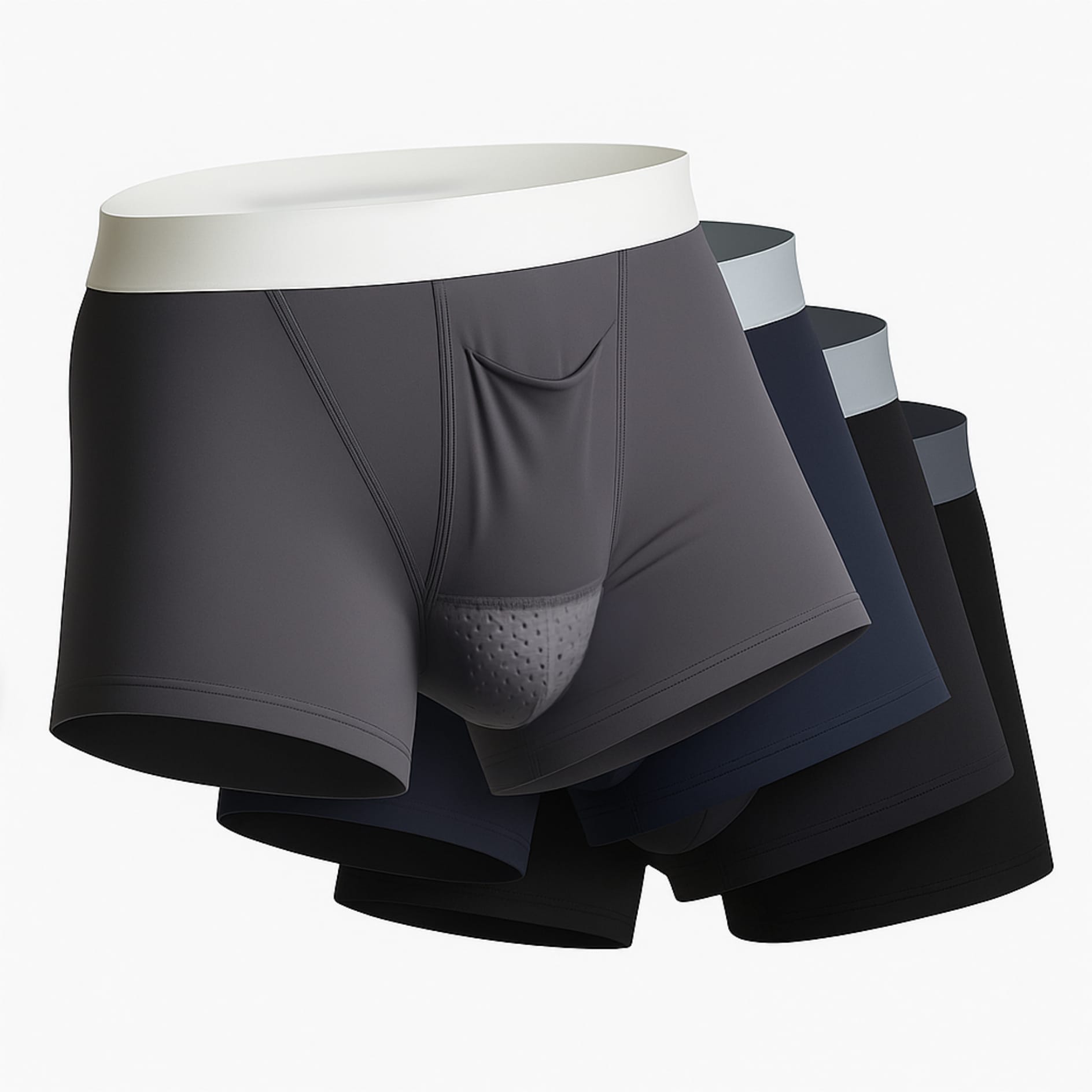
Why Specialized Support Helps
✓ Better support can help reduce the feeling of heaviness.
✓ Breathable, cooling fabrics may help with temperature regulation.
✓ A purpose-built fit can feel more comfortable for daily wear and recovery.
Take the Next Step
Choose Varicocele Support Designed for Cooling, Lift, and Daily Comfort
You’ve seen why regular underwear often falls short. Now view the purpose-built option made to reduce heaviness, improve support, and feel comfortable enough for everyday wear.
✓ Cooling mesh comfort
✓ Supportive ergonomic lift
✓ Discreet for all-day wear