
Experiencing a returning dull ache or persistent swelling after a varicocelectomy or embolization can cause immediate concern. When post-surgical pain resurfaces, confirming whether veins are still dilated requires proper medical imaging. Choosing between an MRI vs ultrasound varicocele scan is a common decision point during a recurrent varicocele diagnosis. Both imaging modalities offer clear views of testicular blood flow, but they differ significantly in cost, accessibility, and practical detail. Understanding how doctors detect persistent veins helps you prepare for follow-up appointments and clear post-surgical imaging.
Why Do Varicoceles Recur? Failed Ligation and Collaterals
A varicocele occurs when valves within the pampiniform plexus (the network of veins draining the testicles) fail to function correctly, allowing blood to pool. Surgery or embolization aims to tie off or block these malfunctioning channels. However, blood vessels sometimes adapt or bypass the treated area.
Understanding why varicoceles can return after surgery involves looking at vascular anatomy. Recurrence generally stems from two main issues:
- Failed Ligation or Occlusion: During open, laparoscopic, or microsurgical procedures, a small collateral vein may be missed. If even one small vessel remains unligated, venous pressure forces blood down that remaining path over time, causing it to dilate.
- Development of Parallel Collateral Veins: The human vascular network is dynamic. When primary veins are closed off, tiny parallel micro-veins can gradually enlarge to take on the workload, recreating the same pooled blood conditions.
- Coil Migration or Recanalization: Patients who undergo catheter-based procedures might experience varicocele recurrence after embolization if metallic coils shift or if the blocked vein forms a new path around the clot.
According to European Association of Urology guidelines, recurrence rates vary between 1% and 15% depending on the initial surgical technique used. Pinpointing the exact cause requires precise diagnostic tools.
Doppler Ultrasound vs. MRI: Which is More Accurate for Recurrence?
When investigating suspected recurrent veins, physicians rely on imaging to assess blood vessel diameter and flow direction during a Valsalva maneuver (bearing down). While color Doppler ultrasound remains the gold standard initial test, magnetic resonance imaging (MRI) offers unique diagnostic advantages in select situations.

Discover specialized varicocele underwear designed for daily comfort, support, and recovery — a more targeted option for men who want support without compromise.
Discover the Recommended OptionEvaluating Doppler ultrasound results allows urologists to observe real-time blood movement. Ultrasound uses sound waves to measure retrograde flow (backflow) and assess vein diameter in both resting and upright positions. Clinical studies indexed on PubMed confirm that high-frequency color Doppler ultrasound achieves high sensitivity and specificity for primary and recurrent venous dilatation.
Scrotal MRI, on the other hand, excels when anatomical pathways are altered by previous scar tissue, or when searching for deep retroperitoneal collateral veins. Magnetic resonance angiography (MRA) can map intricate vascular structures without radiation or operator-dependent ultrasound errors. However, MRI is considerably more expensive and less widely available for routine scrotal evaluations.
| Diagnostic Feature | Color Doppler Ultrasound | Scrotal MRI / MRA |
|---|---|---|
| Primary Benefit | Real-time flow dynamics with Valsalva maneuver | 3D mapping of deep retroperitoneal collaterals |
| Accessibility & Cost | Widely available, lower cost | Requires specialized facility, higher cost |
| Patient Positioning | Dynamic (supine and standing) | Static (supine only) |
| Sensitivity for Recurrence | High (first-line standard) | High (second-line or detailed cases) |
| Effect of Scar Tissue | May cause acoustic shadowing | Unaffected by local surgical scar tissue |
For most men, a Doppler scan for recurrence is sufficient to confirm whether blood is flowing backward into the scrotum. If ultrasound results are inconclusive despite persistent symptoms, a urologist may order an MRI to check for higher retroperitoneal collaterals or unusual vascular anomalies.
Finding Comfort During the Recovery Verification Phase
Waiting for imaging appointments and follow-up reviews can be physically uncomfortable. Gravity continually pulls on the scrotum, putting mechanical stress on sensitive, recovering veins and surrounding nerve pathways. While medical imaging determines the exact anatomical state of your veins, day-to-day symptom management remains essential.
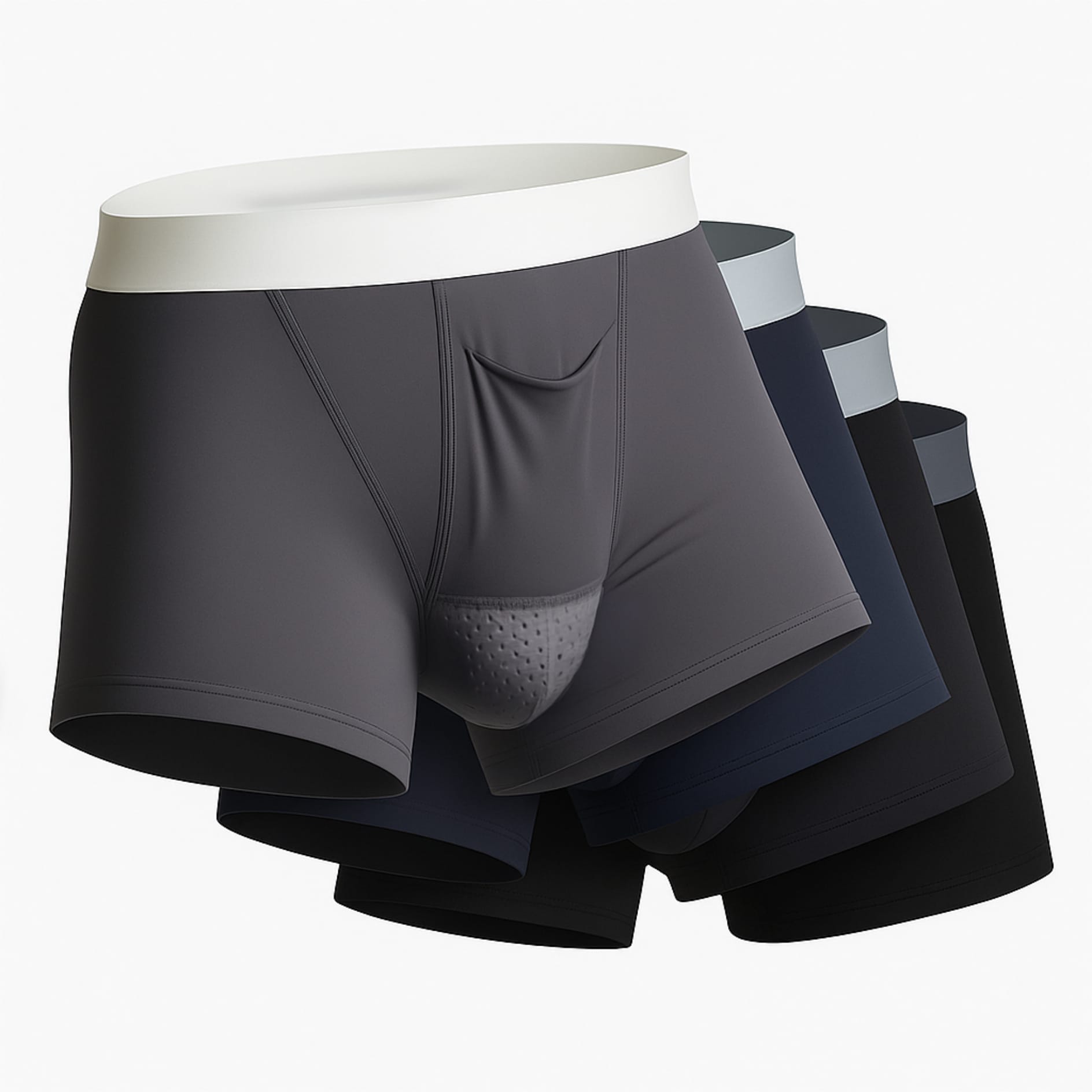
Scrotal support garments do not repair damaged venous valves or cure underlying vascular anatomy. However, elevating the scrotum reduces gravity-induced tension on the spermatic cord. Wearing structured suspension such as Varicocele Underwear with Support & Cooling offers physical comfort and targeted relief from recurring dull aches while you navigate post-surgical diagnostics.
Following a solid post-varicocelectomy recovery guide helps reduce external strain while awaiting clear post-surgical imaging results. Combining supportive garments with rest and temporary activity modification helps maintain everyday comfort.
Frequently Asked Questions
Can Doppler ultrasound miss a recurrent varicocele?
While Doppler ultrasound is highly accurate, it can occasionally miss small or deep collateral veins if the patient is evaluated only while lying flat. Performing the scan in both standing and lying positions with a Valsalva maneuver significantly increases accuracy and prevents missed diagnoses.
Is MRI better than ultrasound for post-surgical imaging?
MRI is not inherently better for every patient, but it offers advantages in involved cases. While ultrasound evaluates dynamic blood flow efficiently at a lower cost, MRI provides detailed three-dimensional mapping of pelvic veins when surgical scarring or deep collaterals obscure ultrasound views.
How long after surgery should imaging be performed to check for recurrence?
Urologists typically recommend waiting at least three to six months after surgery before ordering post-surgical imaging. This timeframe allows post-operative swelling and fluid accumulation to resolve completely, ensuring that diagnostic scans reflect permanent venous flow patterns rather than temporary surgical healing.
Practical Takeaway
Color Doppler ultrasound remains the primary, highly reliable diagnostic tool for detecting recurrent varicoceles due to its ability to measure dynamic blood flow in real time. Reserve scrotal MRI for involved cases where ultrasound results are inconclusive or deep collaterals are suspected. While awaiting medical evaluation, protect yourself from daily mechanical drag by using proper scrotal suspension garments to maintain physical comfort.
Related Articles



Choose Varicocele Support Designed for Cooling, Lift, and Daily Comfort
You’ve seen why regular underwear often falls short. Now view the purpose-built option made to reduce heaviness, improve support, and feel comfortable enough for everyday wear.