
Why Right-Sided Varicoceles Demand Attention
Anatomical asymmetry explains why nearly ninety percent of all varicoceles occur on the left side of the scrotum. The left internal spermatic vein connects into the left renal vein at a perpendicular ninety-degree angle, exposing it to higher backpressure and structural compression between the aorta and superior mesenteric artery. In contrast, the right internal spermatic vein empties directly into the wide, low-resistance inferior vena cava. Understanding why a left side varicocele more common diagnosis is standard helps clarify why isolated right-sided enlargement represents an atypical clinical event. When right spermatic veins dilate, mechanical resistance downstream is preventing blood from reaching the central circulation.Physicians categorize right side vein red flags based on their onset speed and their behavior during physical examination. A classic varicocele typically softens, empties, or fully decompresses when a patient moves from standing upright to lying completely flat on an examination table. If an isolated right varicocele remains firm, engorged, and visibly distended while you are lying flat, this indicates rigid extrinsic vascular compression rather than simple valve failure. In clinical practice, this persistent firmness suggests that an internal anatomical mass is exerting pressure against the vascular wall, holding blood trapped inside the scrotal sac regardless of gravity.The Retroperitoneal Scan Protocol
When an adult patient presents with an isolated or acute right-sided varicocele, standard testicular ultrasound is only the initial step of the diagnostic pathway. Established clinical guidelines for varicocele evaluation instruct clinicians to inspect the entire retroperitoneum, which is the anatomical space located behind the abdominal lining. This diagnostic protocol includes contrast-enhanced computed tomography (CT) of the abdomen and pelvis or high-resolution magnetic resonance imaging (MRI). These cross-sectional imaging tools allow radiologists to track the entire route of the right gonadal vein from the deep inguinal ring directly to its insertion into the inferior vena cava.Ultrasound imaging of the scrotum uses color Doppler to measure retrograde venous flow during resting respiration and forced exhalation maneuvers. However, scrotal imaging alone cannot determine whether an abdominal tumor, swollen lymph node, or vascular anomaly is blocking venous return miles upstream. Investigating the link between a sudden onset varicocele and kidney issues prevents diagnostic delays by pinpointing exact anatomical bottlenecks. Contrast scans clearly delineate kidney margins, check for retroperitoneal lymphadenopathy, and confirm that the interior of the major abdominal veins remains completely open and free of blockages.Ruling Out Kidney Masses and Vena Cava Issues
The primary diagnostic concern during retroperitoneal imaging is ruling out renal cell carcinoma (RCC) and inferior vena cava thrombus. Renal tumors can grow into local vascular branches, occasionally extending an intravascular tumor thrombus directly into the inferior vena cava lumen. This physical occlusion obstructs the opening of the right internal spermatic vein, causing blood to back up into the scrotum under continuous pressure. Other retroperitoneal conditions, such as retroperitoneal fibrosis, benign cysts, adrenal masses, or enlarged lymphatic tissue, can also press against the vena cava from the outside. Reviewing our complete varicocele guide provides additional perspective on how deep abdominal structures intersect with scrotal vascular health.| Clinical Metric | Left-Sided Varicocele | Right-Sided Varicocele (Red Flag) |
|---|---|---|
| Typical Onset | Gradual, commonly during puberty | Sudden onset in adulthood or late adolescence |
| Venous Drainage Route | Left renal vein at a 90-degree angle | Direct drainage into the inferior vena cava |
| Supine Position Response | Decompresses or softens when lying flat | Frequently remains rigid and fully distended |
| Primary Imaging Needed | Scrotal color Doppler ultrasound | Abdominal and pelvic contrast CT or MRI |
| Underlying Risk Profile | Benign venous valve insufficiency | Extrinsic mass, vascular thrombus, or renal growth |
| Immediate Next Step | Symptom control, fertility monitoring | Urgent retroperitoneal and renal imaging |
Related Articles




Expert Recommendation
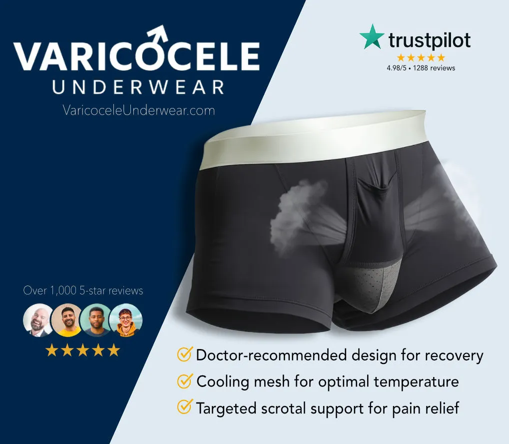
Discover specialized varicocele underwear designed for daily comfort, support, and recovery — a more targeted option for men who want support without compromise.
Discover the Recommended Option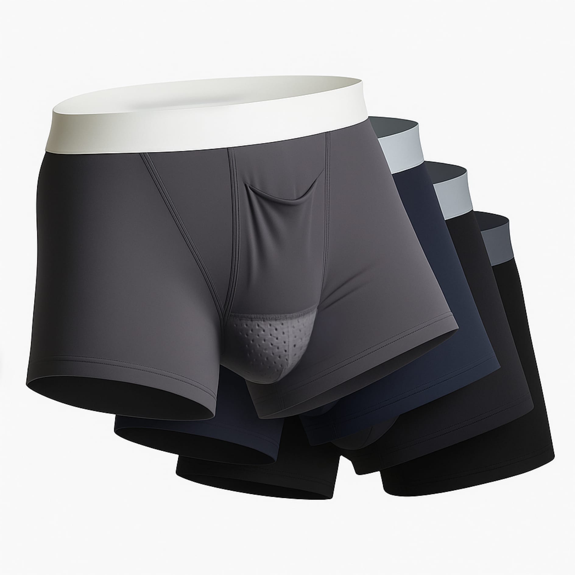
Why Specialized Support Helps
✓ Better support can help reduce the feeling of heaviness.
✓ Breathable, cooling fabrics may help with temperature regulation.
✓ A purpose-built fit can feel more comfortable for daily wear and recovery.
Take the Next Step
Choose Varicocele Support Designed for Cooling, Lift, and Daily Comfort
You’ve seen why regular underwear often falls short. Now view the purpose-built option made to reduce heaviness, improve support, and feel comfortable enough for everyday wear.
✓ Cooling mesh comfort
✓ Supportive ergonomic lift
✓ Discreet for all-day wear