
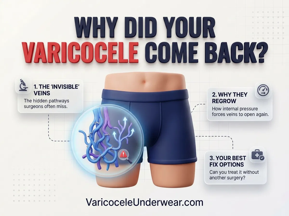
A varicocele recurrence after microsurgery feels like a massive emotional blow, especially when you spent weeks resting and assumed the problem was solved. Subinguinal microsurgical varicocelectomy remains the gold standard procedure with the lowest failure rates in modern urology, usually under two to three percent. Yet, discovering that enlarged scrotal veins have returned or that dull scrotal ache has returned creates immediate panic about fertility, testosterone levels, and comfort. Understanding why veins regrow fix options gives you a clear roadmap forward. When dilated veins reappear, they are rarely the exact same vessels that your surgeon tied off. Instead, recurrence usually involves tiny parallel veins expanding to handle redirected blood flow, or subtle collateral channels that went unnoticed during the original repair. Exploring secondary treatment for recurring veins allows you to regain control and find lasting relief.
Why Varicoceles Can Return After Microsurgery
To understand post-surgical recurrence, you first need to examine how the original operation works. During an operating microscope repair, a urologist creates an incision low in the groin. Using high magnification, the surgeon isolates the spermatic cord, preserves the tiny testicular artery and lymph channels, and ligates (ties off) every visible internal spermatic vein. Closing these faulty pathways forces venous blood to exit the testicle through normal channels that have working one-way valves. However, the human vascular network is dynamic and interconnected. When patients ask can varicocele return after surgery, the answer lies in vascular remodeling. Blood pressure within the pelvis does not simply disappear. If alternative pathways encounter elevated hydrostatic pressure, previously dormant or microscopic vessels gradually stretch, widen, and form a secondary varicocele over several months.
Clinical distinctions between persistence and true recurrence are critical during your follow-up evaluations. True surgical persistence means blood reflux never ceased; the original veins were missed or incompletely ligated, leaving the varicocele palpable immediately after swelling subsides. In contrast, true recurrence occurs after months or years of successful resolution, where new vessels gradually dilate. Following the standard microsurgical varicocelectomy recovery timeline, your urologist typically orders a scrotal color Doppler ultrasound at three to six months. This test confirms whether venous flow is actually reversing downward during a Valsalva maneuver (bearing down) or if you are simply feeling residual, harmless scar tissue known as thrombosis.

Discover specialized varicocele underwear designed for daily comfort, support, and recovery — a more targeted option for men who want support without compromise.
Discover the Recommended OptionIdentifying Missed Veins and Collateral Channels
When a surgeon explores the spermatic cord, they manage a fragile maze of blood vessels. Surgical oversight rarely stems from lack of skill; rather, human vascular anatomy varies drastically between individuals. The pampiniform plexus (the collection of veins cooling the testicle) communicates with external spermatic veins, cremasteric branches, and gubernacular veins that exit from the lower pole of the testis. If a patient possesses retroperitoneal collateral channels branching high up in the abdomen, backward blood flow bypasses the ligation site entirely. These bypass loops redirect pressure back down into the scrotum. Similar structural challenges explain why men also experience varicocele recurrence after embolization when coil placement misses accessory branches.
Gubernacular veins represent one of the most frequent anatomical causes of microsurgical failure. If the urologist does not deliver (elevate) the testicle out of the incision to check the bottom attachment, these lower gubernacular branches remain intact. Over time, gravity and intra-abdominal strain force venous blood down through these untreated pathways. Furthermore, anatomical variants like the nutcracker phenomenon (compression of the left renal vein between the aorta and mesenteric artery) generate high venous backpressure. This force continuously searches for any tiny bypass vessel to dilate, ultimately regenerating scrotal pooling.

Daily conservative care is essential when dealing with secondary vein dilation or awaiting revision procedures. When secondary veins begin stretching, gravity accelerates venous pooling, raising scrotal temperature and straining tender nerves. Wearing post-surgical varicocele recurrence underwear creates targeted, upward scrotal elevation that neutralizes downward hydrostatic drag. This mechanical support stops delicate collateral veins from overstretching while custom cooling architecture dissipates trapped heat to protect your Leydig cells and developing sperm. Integrating the best varicocele underwear into your daily routine provides a reliable baseline of physical comfort during long workdays, heavy lifting, or gym workouts.
| Recurrence Mechanism | Clinical Characteristics | Recommended Secondary Fix |
|---|---|---|
| Missed Gubernacular Veins | Persistent low-scrotal fullness near the bottom pole of the testicle. | Surgical revision with testicular delivery or targeted percutaneous sclerotherapy. |
| External Spermatic Collaterals | Superficial dilation outside the main spermatic cord fascia. | Transvenous coil embolization or cremasteric vein ligation. |
| High Retroperitoneal Bypass | Reflux originates deep above the internal inguinal ring. | Retrograde venography with endovascular coil and foam embolization. |
| Renal Vein Compression | Persistent bilateral or severe left-sided pressure resistant to ligation. | Renal duplex imaging, conservative pelvic decompression, and specialized vascular stenting if symptomatic. |
Revision Surgery and Embolization Options for Recurrence
If recurring veins cause persistent testicular pain, abnormal semen parameters, or progressive scrotal heaviness, revision treatment options are both safe and successful. Modern clinical guidelines, documented extensively in published urological re-intervention literature, frequently recommend percutaneous embolization as the first-line secondary treatment for recurring veins. During embolization, an interventional radiologist inserts a tiny catheter through the femoral or jugular vein under local anesthesia. By injecting contrast dye directly into the venous tree (diagnostic venography), the radiologist identifies the exact rogue collateral branches that bypassed the original surgical ties. Microscopic coils or sclerosant liquids are then deployed directly into those vessels, sealing off the recurrence with minimal downtime.
When embolization is not anatomically feasible, repeat microsurgical varicocelectomy remains a viable path. A revision microsurgical procedure requires exceptional surgical precision because post-operative scar tissue from the initial surgery coats the delicate testicular artery and lymphatic channels. An experienced microsurgeon will often choose an inguinal approach just above the previous subinguinal scar to enter clean tissue planes, clearly isolating the dilated collateral vessels. By combining advanced diagnostic imaging, tailored revision procedures, and consistent scrotal support, men facing varicocele recurrence can successfully eliminate venous reflux, restore natural comfort, and safeguard their long-term reproductive health.
Related Articles

Choose Varicocele Support Designed for Cooling, Lift, and Daily Comfort
You’ve seen why regular underwear often falls short. Now view the purpose-built option made to reduce heaviness, improve support, and feel comfortable enough for everyday wear.